Products
 Products
 Support
 Support


 中文
中文 Eng
Eng


 2013-01-04
 Tutorial
The development of micro technology, so that scientists can more easily penetrate into the micro world. But under the ordinary microscope, the appearance of the cell is the same, it is difficult to distinguish. To this end, scientists have invented a variety of ways: the use of genetic engineering technology to transform the cells, using dye to cell dyeing...... Finally, in view of the microscope, the cell is no longer monotonous, but a beautiful scene.
Whether we like it or not in front of the object, the eyes will always use the same kind of gathering information: retinal cells capture photons. The information will be passed to the brain, the brain reduction for the picture. If the object is too small, the reflection of the photon is too small, the human eye can not see the structure of it. At this time, we need to observe the microscopic technique. This paper shows the pictures, not only has important academic value, more strong artistic beauty. These images represent the most advanced optical microscopy techniques in biological research.
At present, optical microscopy is undergoing an unprecedented change. Scientists use novel fluorescent markers and gene engineering technology modification of tissue samples, let microscope in tissue samples to become colorful, open the door leading to the "discovery". It is a new technology, which is taken by the researchers. By this technique, each mouse brain nerve showing a variety of colors, legible, let us can in the complexity of the neural network tracking analysis of specific axonal, can also draw a complete neural network mapping -- for old imaging technology, it is impossible to finish the task.
The accuracy of the microscope is also improved. We can made a mark in a particular protein, and then use the microscope to observe its activities in the organization line; cell division and differentiation in the process of every detail, also can take in everything in a glance. Researchers can quickly capture in bright light, capturing instantaneous events within a cell or tissue, to observe intracellular fine life process under the weak light. With the development of micro technology, the contradiction between the speed and resolution of image acquisition will be resolved.
At present, several microscopic techniques can even the most subtle biological structures (and treatment were observed in a large number of observation data), the wide application of these techniques, for us to understand the essence of life laid the solid foundation.
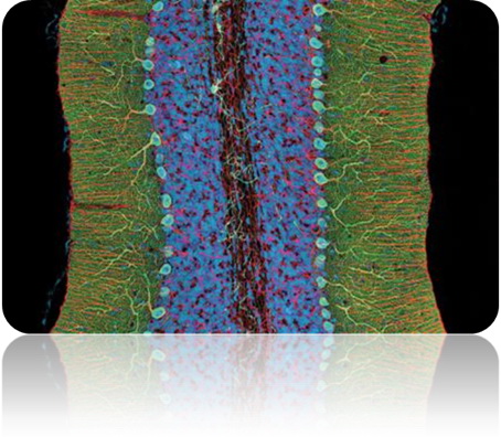
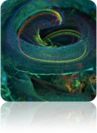
Complex brain
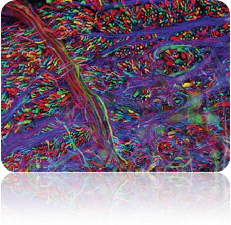
Complex brain
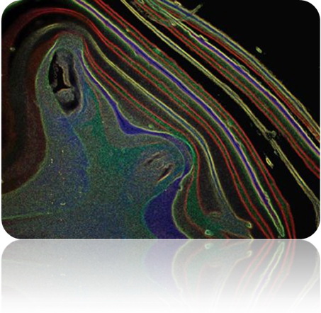
Complex brains: use two-photon microscopy (2-photon microscopy) of the University of California, San Diego, the Thomas deerinck (Thomas Deerinck), shooting to the a piece of only 400 mu m thick mouse cerebellar tissue samples of fine microstructure (pictured above), the green is Purkinje cells (Purkinje neuron), red is astrocytes (glial cell), blue is nucleus. Jean Rivet (Livet Jean), the Harvard University (), using confocal microscopy (microscopy confocal), a genetically engineered mouse brain stem tissue slices (340 m). As a result of the genetic modification, each neuron in the mouse presents a different color (see below). To give neurons a different color (i.e., "Brainbow"), scientists will be able to observe the direction of a single axon in the complex neural network.
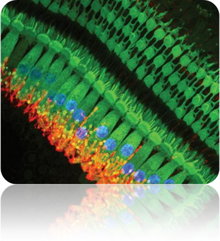
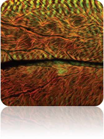
Mouse inner ear hair cells
Tissue structure of mouse inner ear
Because the space is narrow and not easy to separate, inner ear structure is very difficult to observe. University of North Carolina Wilmington campus of Sonia Piot (Sonja pyott) captured the mouse inner ear hair cells (above left), these cells can be mechanical convert sound waves into electrical pulse signal. In the picture, the hair cells are green, and the cells of the hair cells are red and blue, then the nucleus (the confocal microscopy technique). Glenn MacDonald (MacDonald Glen), the University of Washington, uses a similar staining method to capture the tissue structure of the mouse inner ear (confocal microscopy).
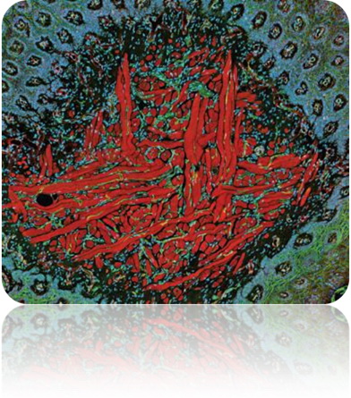
Cross section of mouse tongue muscles
Muscle fiber in Drosophila
Muscle cells constitute a tough muscle tissue. Cross section of the tongue muscles of mice were shown in the image above, by the University of California, San Diego's Thomas deerinck (Thomas Deerinck) shooting. The following picture shows the hand of Hermann Aeberli (Aberle Hermann) of the University of Muenster, Germany, showing the enlarged muscle fibers of the fruit flies. Because of the genetic variation, the muscle fibers of the fruit fly look disorganized (confocal microscopy).
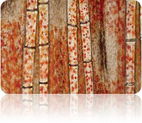
Fish fin bone
Goats bone 4 times
The fins and goat bone: two picture shows, which are dense tissue structure of the vertebrate body. Ramat Gan, Israel, Samuel Silberman Shamuel Silberman put a fish fin bone magnified a hundred times, and there was on top of the mottled autumn (using fiber optic lighting technology). In order to observe the bone formation changes in bone mineral density and mineral content of the increasing degree, the city of Tampa, Florida Mo Moffett cancer center mark Lloyd (Mark Lloyd) and Noel Clark (Noel Clark) the goat bone enlarged four times (see chart, Hirono microscopy).
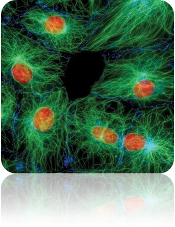
Microtubules in fibroblasts
Microtubules are formed around the chromosomes (blue).
Here is Jan Schmoranza (Sch-moranzer Jan), the Columbia University, the cell membrane of the cells treated by serum starvation, and the structure of the microtubules (green). From the view of the graph, the microtubules of fibroblasts have shown abnormal behavior. The diameter of the microtubules is about 20nm, usually, when there is a gap in the cell membrane, the microtubules will aggregate at the breach, but the situation is not the case. In the interphase cell, Duke U - serdar, Tulu (U. serdar Tulu) in 138 mu m wide horizons captured the chromosome (blue) around is the formation of microtubules (yellow, below).
These pictures, I can not help but think of the famous physicist Richard Feynman (Feynman Richard) in the "fun" in the story of a story. A friend of the Feynman had thought that scientists recognize the beauty of flowers not artists deeply, but also the beautiful flowers open at sixes and sevens, eventually become uninteresting things. Feynman did not agree with the friend's point of view, he said: "I think he is really a bit funny. First of all, what is the difference between him and me and what I see? I believe that even if I do not have the same aesthetic training as he has, but also to appreciate the beauty of a flower...... Let us imagine in cell movement, its perplexing is not a beauty? I mean, the beauty of the flower is not just in the macroscopic form, in the microscopic world, its inner structure is equally fascinating. And flowers to insects of Providence and fight fight Yan, which in itself is a very interesting things, from the side that insects may also be able to distinguish between colors. To see the beautiful flowers, I would like to find out a question: the lower animals also know how to appreciate the beauty of flowers? Why do they have the ability to taste? These interesting questions have proved that scientific knowledge will only make the flowers become more mysterious, more exciting, more awe."